Therapy of Onychomycosis Using a Short-Pulse Nd: YAG Laser: An Example
Dr. Daniel Waldman
Blue Ridge Foot Centers
Intro
Onychomycosis, an ailment induced by dermatophytes, non-dermatophytes and Candida albicans varieties of fungus, is one of the most usual infection of the nail, influencing 2-8 % of the general populace, and enhancing to 12-28 % in adults 60 years or older., Tacks that are contaminated with onychomycosis have yellow or brownish staining, a thickened nail bowl, and crumbling edges.2 These top-notches can easily result in toenail pain, secondary bacterial infections and psychosocial issues such as stress and anxiety, depression, loss of self-confidence, dodging of affection and impaired connections, all which can badly affect a customer's high quality of life., A number of variables featuring reduced blood flow, longer snowballing time of exposure to fungis, nail stress and immune concession such as diabetic issues make individuals a lot more vulnerable to infection. High fees of perseverance and recurrence make onychomycosis incredibly hard to address.
The major therapy approaches for onychomycosis are systemic treatment with the administration of oral antifungal medications and particular antifungal creams administered straight to the contaminated nails. The most typical oral antifungal medicines include terbinafine, fluconazole, and itraconazole.6 Long term treatment rates for dental antifungals vary from 21 % to 53 %. Wide spread treatment requires liver examinations previously, during, and after treatment, and could have significant side effects including looseness of the bowels, dyspepsia, rashes, taste disturbances and stomach ache.6 Client conformity to the blood screening as well as to the demand to stop use of all liquors during the program of therapy can be an obstacle. Particular brokers utilized to address onychomycosis feature nail laquers containing ciclopiroxolamine, amorolfine, toconazole or a combo of these brokers.6 Results from many medical researches indicate that topical antifungal treatment prices could vary from 21 % to 36 %. Variables that make topical antifungals unsuccessful feature lasting application (9-12 months), shortage of patient compliance, major side effects, and failing to deliver the medicine to the nail bed.,.
Laser treatment has recently become a viable therapy option for onychomycosis. Because of its quick therapy period, possibility for efficacy without wide spread therapy or blood monitoring, few contraindications and no significant adverse effects, laser treatment has swiftly come to be a favored treatment alternative for onychomycosis. In addition to these perks, many researches have actually demonstrated that laser treatment is both safe and efficient in boosting the aesthetic look of nails contaminated with onychomycosis.2, 3 This paper goes over the efficacy of the PinPointe FootLaser as a therapy alternative for onychomycosis. To avail of the latest breakthroughs in onychomycosis treatment, kindly follow the link.
Approaches.
Blue Spine Foot Centers has been making use of the PinPointe laser to treat onychomycosis for three years. Before therapy with the laser, customers obtain considerable debridement making use of nippers, curette and a medical drill to eliminate the distal onycholytic nail platter and thinning of the hypertrophic nail platter to 1 mm density. Smoke evacuation is made use of to remove the nail of debridement particles. Patients are then managed with the PinPointe short-pulsed Nd: YAG FootLaser. The laser makes use of the 1064 nm wavelength, 200 millijoules of power, a pulse width of A HUNDRED microseconds, place dimension of 1.5 mm and a power setting of 6 watts. Each place dimension of 1.5 mm obtains 10 micropulses over 0.5 secs and each micropulse delivers 22 Joules/cm2 over 100 microseconds. Laser places were given in a grid design with 1.0-1.5 mm spacing. The managing medical professional holds the handpiece vertical while making makes two passes of the laser, varying between transverse and longitudinally. Each pass around the nail additionally features 2-3 mm around the nail. Hop over here for more info about onychomycosis treatment.
People are given with an appropriate foot treatment regimen to be made use of post-treatment. The anti-fungal medication, Clarus, is administered on and around the nails. The suggestions and also the whole area of the nail are submitted when a week making use of a clean emory board. Tack trimmers are exclusively used by the customer and cleansed with massaging liquor prior to make use of. The people are permitted to make use of Dr's Remedy nail polish but advised not to leave it on for greater than 3-5 days. Shoes, flip-flops and espadrilles are disinfected utilizing either Mycomist sprinkle or Sterishoe devices and all-time low of the shower/tub is cleansed making use of Clorox Clean-Up. In addition, Biotin 2.5 mg a day is used to enhance nails.
Situation Report.
A 31-year-old Caucasian female, taking no medication, presented with onychomycosis of the significant toe. Due to the severity of the infection, the patient obtained 3 passes of the PinPointe laser on each toe, making use of the above specifications. All toes on both feet were handled yet just the significant toes were utilized for analysis. The patient returned for follow-up brows through at months 3, 7 and.10. Photos were taken at standard and at the article treatment follow-up and contrasted. Nail renovation was additionally examined using image analysis.
Planimetry/Image Analysis.
Pre-treatment and follow-up photos were taken of the client's significant toes to examine lesion decrease and understandable nail growth. A professional utilized Adobe Photoshop to obtain regular magnification of the toes along with to map the region of understandable nail and the location of contaminated nail. Pictures were neither increased nor filtered for this reason the margin between clear and contaminated nail was untouched.
To attain uniformity with dimensions and analysis, the following definitions were used:
The pictures were opened in ImageJ software application, a National Institutes of Health (NIH) based program made for image analysis and processing. The program transforms pictures in to their personal pixels to ensure that different regions within the picture can easily be examined. In this study, the technician decided on the clear nail location or the contaminated nail location and the program determined, in pixels, the size of the area. These measurements were at that point utilized to examine the changes in understandable nail region and lesion decrease. Basic discrepancy was calculated to determine the variation that existed from the average. Subjective visual assessments were assessed by the doctor using the International Aesthetic Renovation Scale (GAIS). GAIS is a 5 factor scale to examine the treatment end result of the infected area. Subjects filled in a patient satisfaction set of questions during follow-up visits at 3 and 7 months.
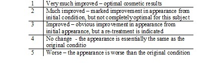
GAIS Examination
Dr. Daniel Waldman
Blue Ridge Foot Centers
Intro
Onychomycosis, an ailment induced by dermatophytes, non-dermatophytes and Candida albicans varieties of fungus, is one of the most usual infection of the nail, influencing 2-8 % of the general populace, and enhancing to 12-28 % in adults 60 years or older., Tacks that are contaminated with onychomycosis have yellow or brownish staining, a thickened nail bowl, and crumbling edges.2 These top-notches can easily result in toenail pain, secondary bacterial infections and psychosocial issues such as stress and anxiety, depression, loss of self-confidence, dodging of affection and impaired connections, all which can badly affect a customer's high quality of life., A number of variables featuring reduced blood flow, longer snowballing time of exposure to fungis, nail stress and immune concession such as diabetic issues make individuals a lot more vulnerable to infection. High fees of perseverance and recurrence make onychomycosis incredibly hard to address.
The major therapy approaches for onychomycosis are systemic treatment with the administration of oral antifungal medications and particular antifungal creams administered straight to the contaminated nails. The most typical oral antifungal medicines include terbinafine, fluconazole, and itraconazole.6 Long term treatment rates for dental antifungals vary from 21 % to 53 %. Wide spread treatment requires liver examinations previously, during, and after treatment, and could have significant side effects including looseness of the bowels, dyspepsia, rashes, taste disturbances and stomach ache.6 Client conformity to the blood screening as well as to the demand to stop use of all liquors during the program of therapy can be an obstacle. Particular brokers utilized to address onychomycosis feature nail laquers containing ciclopiroxolamine, amorolfine, toconazole or a combo of these brokers.6 Results from many medical researches indicate that topical antifungal treatment prices could vary from 21 % to 36 %. Variables that make topical antifungals unsuccessful feature lasting application (9-12 months), shortage of patient compliance, major side effects, and failing to deliver the medicine to the nail bed.,.
Laser treatment has recently become a viable therapy option for onychomycosis. Because of its quick therapy period, possibility for efficacy without wide spread therapy or blood monitoring, few contraindications and no significant adverse effects, laser treatment has swiftly come to be a favored treatment alternative for onychomycosis. In addition to these perks, many researches have actually demonstrated that laser treatment is both safe and efficient in boosting the aesthetic look of nails contaminated with onychomycosis.2, 3 This paper goes over the efficacy of the PinPointe FootLaser as a therapy alternative for onychomycosis. To avail of the latest breakthroughs in onychomycosis treatment, kindly follow the link.
Approaches.
Blue Spine Foot Centers has been making use of the PinPointe laser to treat onychomycosis for three years. Before therapy with the laser, customers obtain considerable debridement making use of nippers, curette and a medical drill to eliminate the distal onycholytic nail platter and thinning of the hypertrophic nail platter to 1 mm density. Smoke evacuation is made use of to remove the nail of debridement particles. Patients are then managed with the PinPointe short-pulsed Nd: YAG FootLaser. The laser makes use of the 1064 nm wavelength, 200 millijoules of power, a pulse width of A HUNDRED microseconds, place dimension of 1.5 mm and a power setting of 6 watts. Each place dimension of 1.5 mm obtains 10 micropulses over 0.5 secs and each micropulse delivers 22 Joules/cm2 over 100 microseconds. Laser places were given in a grid design with 1.0-1.5 mm spacing. The managing medical professional holds the handpiece vertical while making makes two passes of the laser, varying between transverse and longitudinally. Each pass around the nail additionally features 2-3 mm around the nail. Hop over here for more info about onychomycosis treatment.
People are given with an appropriate foot treatment regimen to be made use of post-treatment. The anti-fungal medication, Clarus, is administered on and around the nails. The suggestions and also the whole area of the nail are submitted when a week making use of a clean emory board. Tack trimmers are exclusively used by the customer and cleansed with massaging liquor prior to make use of. The people are permitted to make use of Dr's Remedy nail polish but advised not to leave it on for greater than 3-5 days. Shoes, flip-flops and espadrilles are disinfected utilizing either Mycomist sprinkle or Sterishoe devices and all-time low of the shower/tub is cleansed making use of Clorox Clean-Up. In addition, Biotin 2.5 mg a day is used to enhance nails.
Situation Report.
A 31-year-old Caucasian female, taking no medication, presented with onychomycosis of the significant toe. Due to the severity of the infection, the patient obtained 3 passes of the PinPointe laser on each toe, making use of the above specifications. All toes on both feet were handled yet just the significant toes were utilized for analysis. The patient returned for follow-up brows through at months 3, 7 and.10. Photos were taken at standard and at the article treatment follow-up and contrasted. Nail renovation was additionally examined using image analysis.
Planimetry/Image Analysis.
Pre-treatment and follow-up photos were taken of the client's significant toes to examine lesion decrease and understandable nail growth. A professional utilized Adobe Photoshop to obtain regular magnification of the toes along with to map the region of understandable nail and the location of contaminated nail. Pictures were neither increased nor filtered for this reason the margin between clear and contaminated nail was untouched.
To attain uniformity with dimensions and analysis, the following definitions were used:
- Clear Nail: Uniform in color (pink, red, flesh, or pale) with hassle-free surface and typical density.
- Sick Nail: Irregular in color scheme (milk white, black, brownish, yellow, green) with flakey, altered, roughened, ridges, or thickened nail bowl that might be onycholytic.
- Sore Area: Complete contaminated area featuring staining, thickening and yellowing of nail.
- Sore Location at Follow-up: Overall infected location at follow-up.
- Clear Nail Growth: New understandable nail that has expanded (%) in comparison to the Baseline Clear Nail Location.
- Outright Clear Nail: The baseline lesion location (%) minus the follow-up sore area (%).
- Lesion Reduction (% baseline): The Absolute Lesion Decline % broken down by the Standard Sore Location (%).
The pictures were opened in ImageJ software application, a National Institutes of Health (NIH) based program made for image analysis and processing. The program transforms pictures in to their personal pixels to ensure that different regions within the picture can easily be examined. In this study, the technician decided on the clear nail location or the contaminated nail location and the program determined, in pixels, the size of the area. These measurements were at that point utilized to examine the changes in understandable nail region and lesion decrease. Basic discrepancy was calculated to determine the variation that existed from the average. Subjective visual assessments were assessed by the doctor using the International Aesthetic Renovation Scale (GAIS). GAIS is a 5 factor scale to examine the treatment end result of the infected area. Subjects filled in a patient satisfaction set of questions during follow-up visits at 3 and 7 months.
GAIS Examination
Results
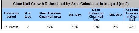
The patient’s two big toes were measured by planimetry and showed a significant increase in clear nail growth and lesion reduction (Figure 1,2). The mean baseline clear nail area for the patient’s two toes was 17% (±32%) of the nail. After 14 months post-treatment, the mean clear nail area was increased to 49% (±5%) of the nail. Using these two measurements, the absolute increase in clear nail was calculated to be 32%. (Table 1).
Figure 1. Patient’s right foot
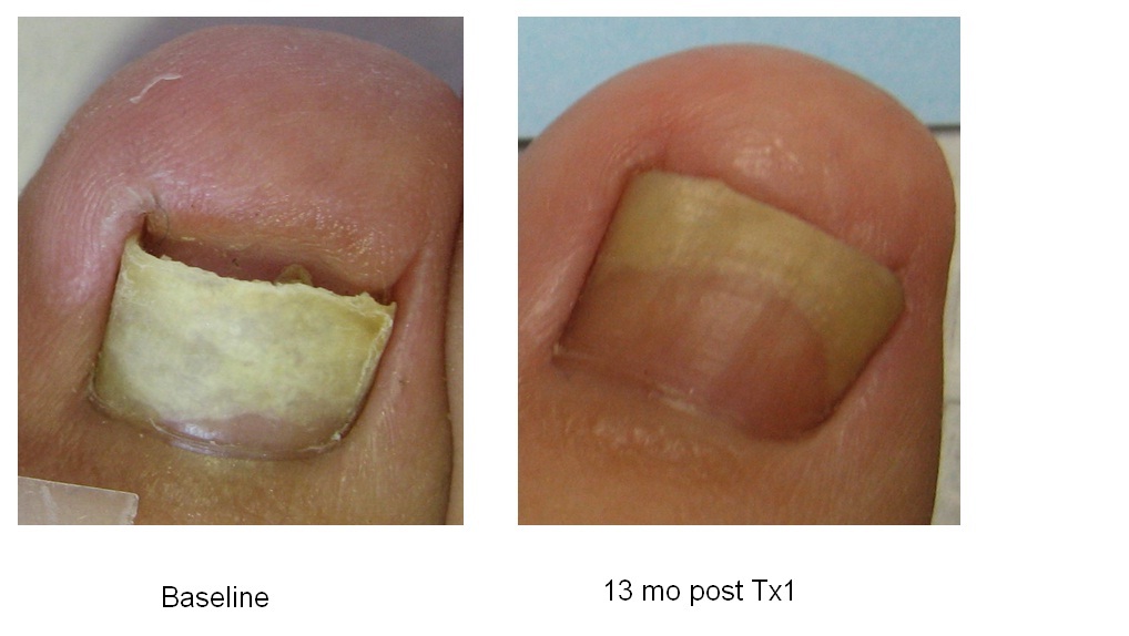
Figure 2. Patient’s Left Foot
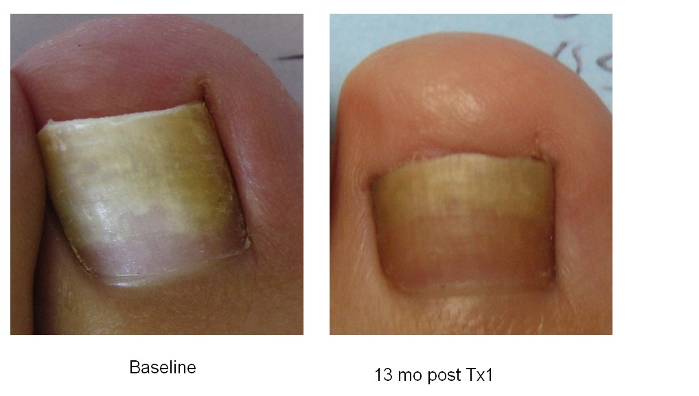
Table 1. Improvement is quantified by increase in clear nail
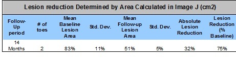
The mean lesion area of the patient’s two big toes was determined to be 83% (±11%) of the nail and was reduced to 51% (±5%) of the nail at 14 months follow up. Based on the mean baseline lesion area and the mean follow up lesion area, absolute lesion reduction was calculated to be 32% and lesion reduction vs. baseline was calculated to be 75%. (Table 2). The physician rated the patient’s clinical outcome a 4 on the Global Aesthetic Improvement Scale.
Table 2. Improvement is quantified as the lesion area reduction (% baseline)
Table 2. Improvement is quantified as the lesion area reduction (% baseline)
Discussion
In our experience, treatment with the PinPointe laser not only allows for significant growth of clear nail in toes infected with onychomycosis but the laser treatment also produces high patient satisfaction. In this case study, the efficacy of the 1064 nm short pulsed Nd:YAG laser was evaluated based on clear nail growth and reduced lesion area. The patient experienced a significant improvement in both areas. The clear nail area of the patient increased from 17% of the nail to 49% of the nail, and there was a 75% reduction in lesion area. In addition to the significant efficacy of the PinPointe Laser treatment as demonstrated by planimetry measurements, the physician also noticed a significant improvement in the nail. Using the Global Aesthetic Improvement Scale, the physician rated the patient’s improvement as a 4, indicating that the nails were much improved and there was marked improvement in appearance from the initial condition. The results of this case study suggest that the 1064 nm Nd:YAG PinPointe Laser could be an effective treatment option for onychomycosis.
Conclusion
The PinPointe laser is an effective treatment option that produces high patient and physician satisfaction. Laser treatment allows for clear nail growth without the significant side effects and complications that come with other treatment modalities.
References
[1] Finch J, Warshaw E. Toenail onychomycosis: current and future treatment options. J Dermatol Ther. 2007; 20:31-46
[1] Hochman L.G. Laser Treatment of onychomycosis using a novel 0.65-millisecond pulsed Nd:YAg 1064-nm laser. Journal of Cosmetic and Laser Therapy. 2011; 13: 2-5
[1] Menevitch Z., Lev D., Hochberg., Palhan M., Lewis A., Enk C.D. Direct Antifungal effect of Femtosecond Laser on Trichophyton rubrum Onchomycosis. Photochemistry and Photobiology. 2010; 86: 476-479.
[1] Finch, J. J., Warshaw E.M. Toenail Onychomycosis: current and future treatment options. Dermatologic Therapy. 2007; 20: 31-46.
[1] Nasir A., Goldstein B., van Cleff, M., Swick L. Clinical Evaluation of Safety and Efficacy of a New Topical treatment for Onychomycosis. Journal of Drugs in Dermatology. October 2011; 10(10): 1186-1191.
[1] Welsh O, Vera-Cabrera L, Welsh E. Onychomycosis. Clinics in Dermatology 2010; 28: 151-159.
[1] Sigurgeirsson, B., J.H. Olafsson, J. B. Steinsson, C. Paul, S. Billstein and E. G. Evans (2002) Long-term effectiveness of treatment with terbinafine vs itraconazole in onchomycosis: A 5-year blinded prospective follow-up study. Arch Dermatol. 138, 353-357
[1] Gupta AK, Schouten JR, Lynch LE. Ciclopirox nail lacquer 8% for the treatment of onychomycosis: A Canadian perspective. Skin therapy Lett 2005;10:1-3
[1] Lesher JL Jr (1999) Oral therapy of common superficial fungal infections of the skin. J Am Acad Dermatol 40(6 pt 2):S31-S34
[1] Koehler AM, Maibach HI (2001) Electronic monitoring in medication adherence measurement. Implications for dermatology. Am J Clin Dermatol 2:7-12
[1] Piraccini BM, Rech G, Tosti A. Photodynamic therapy of onychomycosis caused by Trichophyton rubrum. J Am Acad Dermatol 2008; 59(% Suppl):S75-76.
[1] National Institute of Health
